Main Content
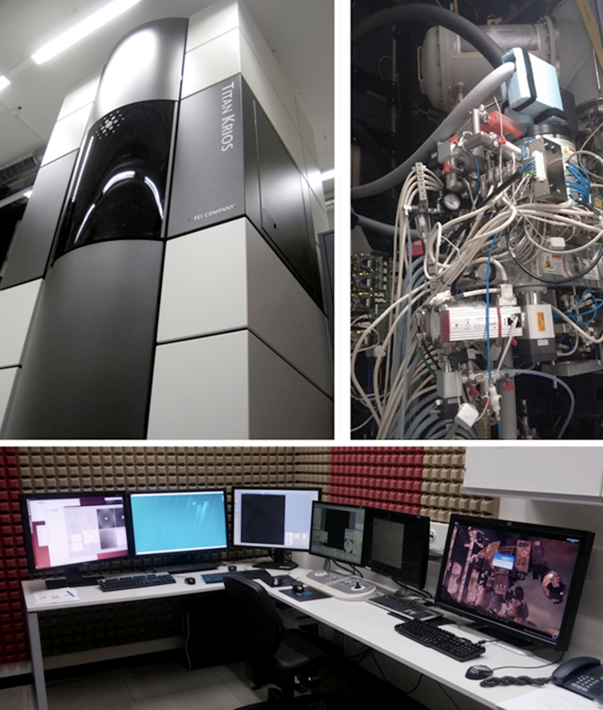
FEI Titan Krios TEM (300kV)
Configuration
80-300kV, FEG. 12-grid liquid nitrogen-cooled stage, Gatan Quantum-LS Energy Filter (GIF) with a Gatan K2 Summit direct electron detector. The instrument is housed within an environmental enclosure to reduce thermal and acoustic influences and increase stability. Online drift-correction and pre-processing capabilities are installed on adjacent computers directly next to the console of the instrument. SerialEM is generally used for automated image acquisition.
Application
The Titan Krios is a high-end transmission electron microscope tailored for molecular and cellular imaging at cryo-temperatures. The high stability permits a full range of applications, including, single particle, helical filament analysis, and electron tomography at ultimate resolution. Cellular imaging of frozen-hydrated cell organelles and cells prepared under cryo-conditions allows observation of specimens while the integrity in their native state is maintained. Applying an energy filter allows zero loss imaging to exclude inelastically scattered electrons. This can improve both contrast and resolution of an image.
Titan Krios Specifications
Accelerating Voltage | 300 kV |
Electron Source | Schottky S-FEG |
Stage Tilt Range | ± 70° |
C1 Apertures | 2000, 70, 50 and 30 µm |
C2 Apertures | 150, 100, 70 and 50 µm |
Objective Apertures | 100, 70 µm |
Operating Temperature | ~80 K |
Information Limit at 300kV at 0° / ±70° tilt | 0.14 nm and 0.23 nm |
Cs | 2.7 mm |
Number of cartridge positions for grids | 12 total |
Sample exchange time | ~3 mins from autoloader cassette to column |
Imaging filter and detector | GIF Quantum LS image filter fitted with a retractable K2 Direct Electron Detector; · Sensor size 3840 x 3712 pixels (14 megapixels) · Sensor active area 19.2 mm x 18.6 mm · Physical pixel size 5 µm · Sensor read out 400 full fps · Transfer speed to computer 40 full fps · DQE >0.5 at 0.5 physical Nyquist · Super-resolution with sub-pixel accuracy for 57 MP effective resolution · Counts individual image electrons (counting mode) |
Acquisition software | SerialEM for single particle and tomography |


