
Main Content
The molecular architecture of cells
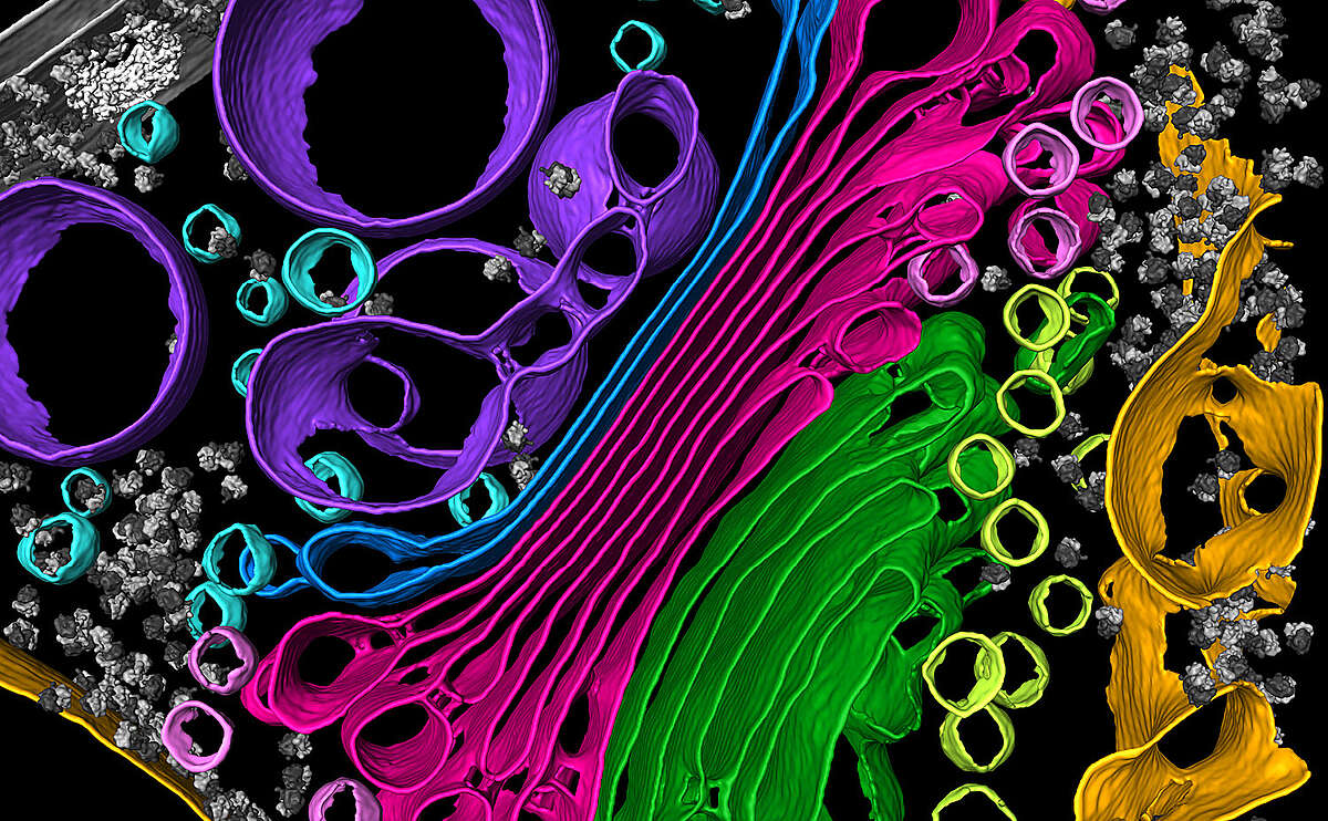
We use cryo-electron tomography to visualize molecular complexes inside native cells, providing new insights into how organelles are built.

Cells perform specific biochemical reactions by concentrating their proteins into subcellular compartments called organelles. These include the nucleus, endoplasmic reticulum, Golgi, mitochondria, chloroplasts, cilia, and phase-separated compartments.
We explore the relationship between organelle form and function by investigating how macromolecules sculpt cellular organelles, and reciprocally, how the shape of each organelle directs the function of its resident macromolecules. Cryo-electron tomography (cryo-ET) provides a window into this new world of “structural cell biology”.
Photosynthesis turns light into life
The process of photosynthesis is essential to life on Earth because it provides food, while replacing atmospheric carbon dioxide with oxygen. A major focus of our research is how the molecular architecture inside chloroplasts and cyanobacteria enables cells to harvest light energy and fix carbon dioxide.
Marine algae in a changing world
In addition to classical model organisms, we study photosynthesis in diverse marine algae, which perform a major portion of the Earth’s carbon fixation and help sustain the Ocean’s biodiversity. We seek to understand how photosynthetic organelles have evolved to the marine environment and how they are affected by environmental stresses caused by climate change.
Cilia are the cell’s antenna
Our lab also studies cilia, hair-like organelles that extend from many unicellular organisms, as well as nearly every cell in the human body. Cilia perform a variety of motility and signaling functions, and thus, cilia dysfunction causes many human diseases. We are interested in understanding the structure and function of cilia across evolution.

