
Throughout our body there is a dense, widely ramified network of blood vessels. It is estimated to be about 150,000 kilometers in length. The vessels supply all cells, even those in most far away regions of the body with nutrients and oxygen and remove the metabolites. Already, very early in embryo development the heart starts to beat and the first blood vessels begin to form. The crux is, that the blood vessels form while blood is flowing through them and their integrity needs to be maintained to prevent blood leakage.
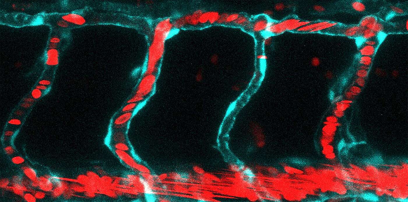
During the development of blood vessels, the vascular cells form an intercellular space into which the blood flows, thus slowly expanding the lumen. In order to withstand this pressure, the cell-cell junctions must remain tight and stable. Together with researchers at Amsterdam UMC, the team led by Prof. Markus Affolter at the Biozentrum, University of Basel, has now been able to demonstrate in zebrafish that the protein Vinculin selectively strengthens specific vascular cell junctions exposed to high pressure and protects them from tearing, particularly in the early stages of vessel formation.
Contact points under high pressure produce “fingers”
As new blood vessels sprout; the vascular cells are initially arranged in a row. They then gradually move over each other and finally align themselves so that an internal tube is formed between them. During the process, they must maintain the contact with their neighboring cells. While the vascular cells form a lumen, the blood surging into it exerts a high pressure, which generates tension on many of the cell junctions. At these sites, tiniest junctional “fingers” emerge, which reinforce the junctions and make them more robust.
The protein Vinculin is required for “finger” formation
“The protein Vinculin accumulates at the cell junctions exposed to great tension from the blood pressure,” explains the head of the study Dr. Heinz-Georg Belting. “It fortifies and stabilizes the junction by causing invagination of the cell membrane forming typical finger-like structures. Vinculin is the driving force for junction reinforcement. If it is lacking, no fingers are produced despite high junctional tension.” Once the lumen is completely open and the blood flow is established, the tension on the specific vascular junctions is relieved and the fingers regress over time.
Mechanical forces influence vessel formation
The role played by blood pressure and flow as well as shear stress particularly in blood vessels formation has still not been resolved in detail. “Here we have a new system that allows us to take a deeper look at how the vascular cells deal with the various mechanical forces during angiogenesis,” says Maria Kotini, first author of the study. In order to further elucidate blood vessel formation, the researchers next want to investigate how large the forces at the cell junctions are, how the force is generally distributed and which further factors activate the formation of junctional fingers.
Original article:
Maria P. Kotini, Miesje M. van der Stoel, Jianmin Yin, Mitchell K. Han, Bettina Kirchmaier, Johan de Rooij, Markus Affolter, Stephan Huveneers and Heinz-Georg Belting. Vinculin controls endothelial cell junction dynamics during vascular lumen formation. Cell Reports, published online 12 April 2022
Contact: Communications, Katrin Bühler