Parkinson’s disease is one of the most common neurodegenerative diseases worldwide. This disease is typically accompanied by motor defects such as the tremor of arms and legs, slowness of movements and muscle rigidity, which occur together with other non-motor symptoms. A characteristic of this progressively worsening and currently unstoppable disease are neuronal inclusions, so called Lewy bodies, that occur in many regions of the brain in the course of the disease. For decades, it was assumed that Parkinson’s disease is caused by deposits of insoluble fibrils consisting of the protein alpha-synculein in the Lewy bodies.
Parkinson inclusions filled with membrane fragments instead of protein fibrils
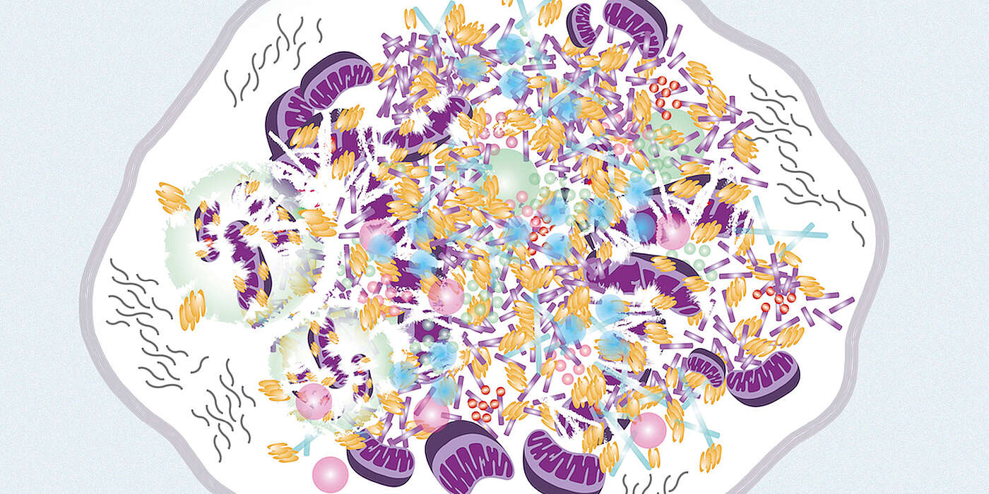
In their current study, the Dutch, German and Swiss researchers, including Prof. Henning Stahlberg’s team, refute this long held common thesis. Using state-of-the-art electron microscopes, they have been able to show that the Lewy bodies contain mainly membrane fragments, lipids and other cellular material instead of the anticipated fibrils.
“We used correlative light and electron microscopy and other advanced light microscopy methods to take a closer look at the brain of deceased Parkinson’s patients and discovered that the Lewy bodies consist mainly of membrane fragments from mitochondria and other organelles, but have in most cases no or only negligible quantities of protein fibrils,” says Stahlberg. “The discovery that alpha-synuclein did not present in the form of fibrils was unexpected for us and the entire research field.”
Currently, the researchers do not know yet where and in what form the protein alpha-synuclein is hidden amongst the membrane fragments and how it is involved in the formation of Lewy bodies. However, their work indicates that the laboratory-based model of alpha-synuclein fibrils as a cause and mechanism of Parkinson’s disease should be revisited. “Our finding indicates that in order to uncover the causes of a disease one needs to be more strongly guided by the exploration of the pathology in humans,” explains Stahlberg.
Ultrastructural insights into cell organelles
“The questions why it has taken so long to better characterize Lewy bodies, can perhaps be answered with the previous sample preparation and electron microscopy methods. Today’s technologies enable us to have a much more detailed look into the morphology of human brain,” explains Stahlberg. “The big question for us now is: How does alpha-synuclein contribute to the formation of Lewy bodies, if not present in form of fibrils?" With their work, the researchers raise many new questions regarding the role of the Lewy bodies in the etiology of Parkinson’s disease. The insights into such intracellular structures also provide important clues for potential therapeutic approaches to prevent or stop the formation and propagation of Lewy pathology in the brain.
Original publication:
Sarah H. Shahmoradian, Amanda J. Lewis, Christel Genoud, Juergen Hench, Tim Moors, Paula Perez-Navarro, Daniel Castano-Diez, Gabriel Schweighauser, Alexandra Graff-Meyer, Kenneth N. Goldie, Rosmarie Suetterlin, Evelien Huisman, Angela Ingrassia, Yvonne de Gier, Annemieke J.M. Rozemuller, Jing Wang, Anne Da Paepe, Johannes Erny, Andreas Staempfli, Joerg Hoernschemeyer, Frederik Grosserueschkamp, Daniel Niedieker, Samir F. El-Mashtoly, Marialuida Quadri, Wilfred F.J. van IJcken, Vincenzo Bonifati, Klaus Gerwert, Bernd Bohrmann, Stephan Frank, Markus Britschgi, Henning Stahlberg, Wilma van de Berg, Matthias E. Lauer. Lewy pathology in Parkinson's disease consists of crowded organelles and lipid membranes. Nature Neuroscience. published online June 24, 2019.
Contact: Communications, Katrin Bühler


